Differential diagnosis
Arterial ulcers are usually found on the toes, feet, lateral aspect or pretibial aspect of lower leg. The ulcer is usually punched out and sharply demarcated. It can be very painful, small and deep with low exudate levels or dry and gangrene may also be present. There are usually weak or absent foot pulses. The surrounding skin may be dry and shiny. These account for around 10% of all ulcers.
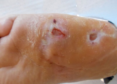
Diabetic foot ulcer. These are usually found on pressure bearing areas, such as soles of feet and the foot margins, commonly on the first or fifth metatarsal head. The ulcer may not be painful if there is accompanied sensory loss due to neuropathy. The ulcer depth will be variable although it may involve tendons or bone and there may be a sinus present. The neuropathic foot will feel warm to touch, and there is often the presence of callous. The neuroischaemic foot may be cool to touch and foot pulses will be absent.

Mixed aetiology. This is a venous leg ulcer with arterial disease in the limb. The patient will have a low ABPI and will exhibit a mixture of signs and symptoms. These account for around 20% of all leg ulcers.
Vasculitis. This is an auto-immune condition which causes healthy blood vessels to become swollen and narrowed it can also be drug induced. Examples are Lupus Erythematosus, Sjogren’s Syndrome, Nodular Arteritis or Wegener’s Granulomatosis.
Other causes. Metabolic, Haematologic, Neoplasia, Infectious, Ulcerating skin disease, Genetic diseases and drug induced are other causes of leg ulcerations.
Further Reading:
NICE clinical knowledge summary on venous leg ulcers

